Skeletal and Muscular System Imaging
Bones, joints, muscles, ligaments, tendons, the spine, and muscles are evaluated in detail for systemic diseases, traumas, cancers, arthritis, and arthrosis-related conditions.
Main Techniques for Evaluation of These Structures:
Arthrography
This technique involves the evaluation of all skeletal and muscular system structures, particularly joints, using X-rays (radiography), fluoroscopy, and other methods to assess pain, restricted movement, and trauma. When additional information is required, MRI and/or CT can be utilized.
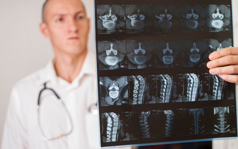
Myelography
A specialized MRI imaging technique that provides detailed visualization of the spinal cord and nerve roots. It is a crucial method for diagnosing and planning the treatment of abnormalities, tumors, herniations, and other pathologies in the spinal canal.
Digital X-ray (Radiography)
A conventional and cost-effective method useful for imaging in this area.
Bone Density Measurement (DEXA)
Measures the mineral density of the spine and hip bones using DEXA (dual-energy absorptiometry). It is performed to diagnose and treat conditions that increase bone mineral loss, such as osteoporosis, before fractures occur, thereby preventing potential fractures. Regular assessments as recommended by your physician help maintain quality of life by preventing fractures.