Magnetic Resonance Imaging (MRI) (3TESLA)
What is MRI (Magnetic Resonance Imaging) / 3-Tesla MRI?
MRI (Magnetic Resonance Imaging) / 3-Tesla MRI is an advanced medical imaging method that obtains high-resolution images of the body through a powerful magnetic field (magnet), radio waves, and advanced computers with specialized software. It is a non-invasive, painless imaging technique that does not require hospitalization. Unlike direct X-ray images (radiographs) and computed tomography (CT), MRI does not use ionizing radiation, making it a patient- and doctor-friendly method. In the near future, revolutionary developments are expected with AI-supported MRI scans, leading to faster imaging, reduced workload, and increased efficiency. AI and machine learning show promise in scan planning, obtaining multidimensional data, supporting clinical interpretation of images, and personalized imaging.
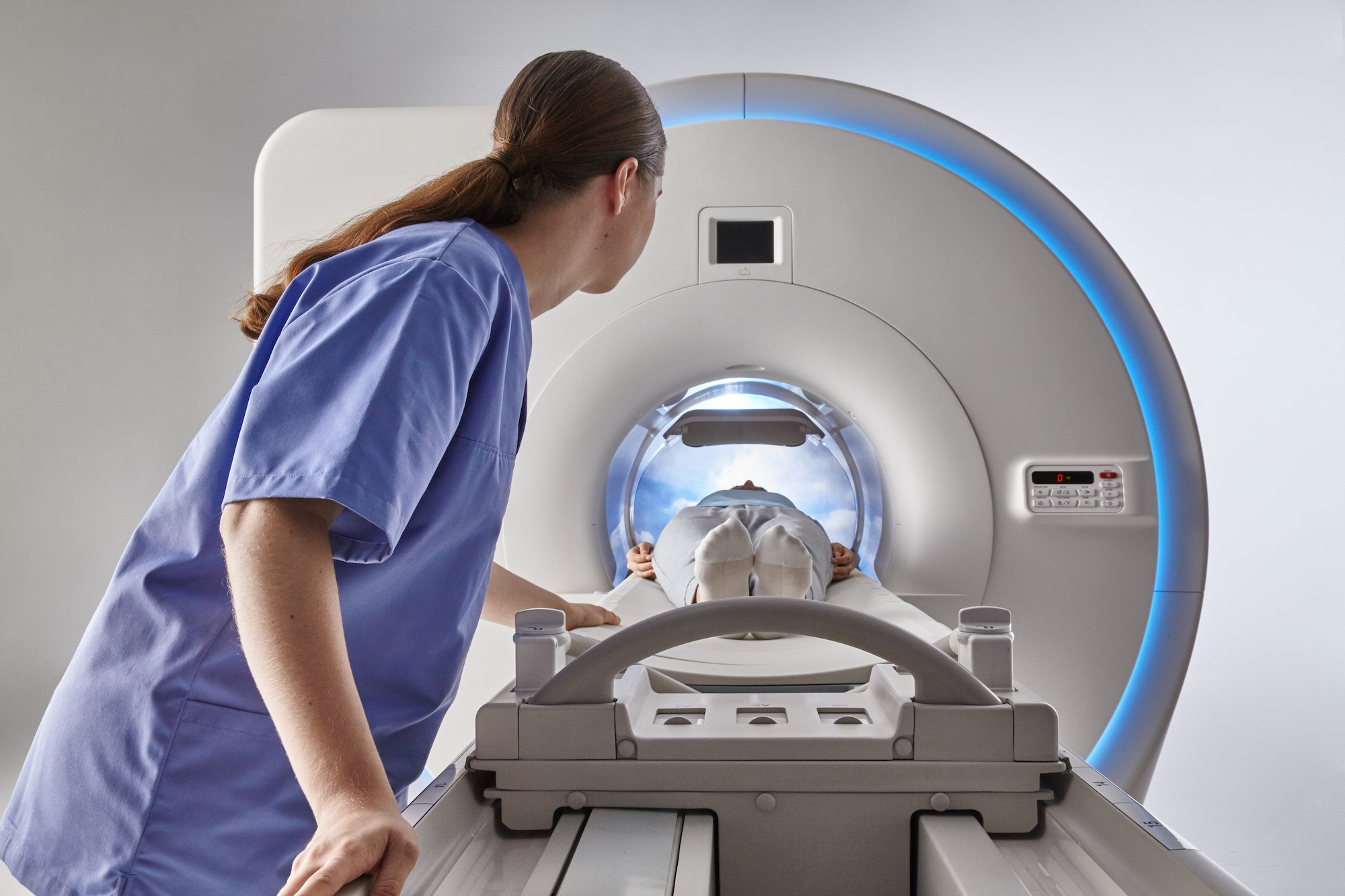
An MRI machine is placed in a specially equipped scanning room and contains very powerful magnets in a cylindrical tunnel-like tube open at both ends. The highest resolution image quality is obtained at the center of the magnet; therefore, the body area to be examined must be positioned inside the tube. For this purpose, when the scan begins, the patient lies on a table that moves slowly to position the area of interest at the center. To obtain clear and high-quality images, it is essential for the patient to remain still and maintain a fixed position during the scan. Belt-like stabilizers may be used for this purpose.
MRI allows examination of nearly the entire body. Images obtained through computers and specialized advanced software provide essential information for disease screening, diagnosis, treatment planning, and evaluating the effectiveness of previous treatments. Since ionizing radiation (X-rays) is not used, it is a prominent imaging method for diseases that require frequent monitoring.
- The MRI scan duration depends on the size of the area to be imaged and the number of slices to be taken, typically ranging from approximately 15 to 90 minutes. Whole-body MRI scans may take longer. During the scan, you generally do not need to hold your breath, making it advantageous for patients with mobility issues, dementia, hearing impairment, etc.
- MRI-guided biopsy and other interventional procedures can also be performed. Thanks to the features of our devices, the images obtained are not limited to horizontal slices; three-dimensional (3-D) images can also be obtained, allowing precise determination of lesion locations. This feature is important for biopsy and surgical planning.
- If brighter and sharper images are required, contrast material may need to be injected intravenously (IV). For this, a catheter is placed in your vein by a doctor, technician, or nurse. This decision is made by your referring physician or the radiologist evaluating the results.
- As the magnetic field strength increases in MRI scans, the frequency difference between fat and water in the body widens. This feature allows for special imaging techniques such as MR spectroscopy, functional MRI, diffusion MRI, and arterial spin labeling.
- MRI is particularly superior in neuroradiology, i.e., imaging and evaluating brain and nerve tissue functions compared to other methods. Perfusion MRI, performed without contrast material using the arterial spin labeling method, provides information about brain perfusion. Since contrast material is not required, this imaging method is advantageous for pediatric patients. In addition to brain perfusion, perfusion MRI can assess the perfusion of most tissues or areas of interest, including the heart.
- By increasing resolution, the chances of detecting very small lesions in cartilage, bone, tendons, and ligaments in the musculoskeletal system are improved. MR neurography and plexus examinations can be successfully performed.
- 3-Tesla MRI devices enable high-resolution imaging of contrast-enhanced and non-contrast MRI angiography.
- It facilitates the application of the most current and accurate prostate diagnostic methods, such as multiparametric prostate imaging and fusion prostate biopsy.
- High signal strength contributes to early diagnosis even in very small lesions in liver and breast diseases.
- Although rare, fetal MRI can also be performed if deemed necessary by your physician.
- In addition to all the special imaging methods mentioned above, 3-Tesla MRI is an indispensable advanced imaging method for detailed evaluation of the gallbladder and bile ducts, kidneys, spleen, pancreas, adrenal glands, uterus, ovaries, blood vessels, lymph nodes, and many other organs and tissues.
- Our institutions (Nişantaşı Centermed, Centermed Akademi, Centermed Plus, and Betemar) are equipped with four state-of-the-art 3-Tesla MRI machines with advanced software and hardware, suitable for high-weight patients, allowing us to meet all requirements in this field on the same day.
Is MRI risky?
MRI scans are not risky, but since this imaging method uses a very powerful magnetic field, it applies a strong magnetic force on iron, steel, and other metal materials in the scanning room, which could rapidly pull objects like a wheelchair into the machine. Extreme caution is required in this situation. Experienced MRI technicians and assistants will inform you about this issue and ensure necessary precautions are taken before you enter the room.
How to prepare for an MRI scan?
- First, ask your physician who requested the MRI to explain why the test is needed, its possible risks, and answer all your questions.
- Before most MRI scans, you can drink water, eat, and take your medications. If a different situation applies, you will be informed in advance when you make your appointment.
- On the day of your appointment, wear comfortable clothing without metal zippers, hooks, or buttons. You may need to undress and wear a single-use gown provided by our institution, suitable for the scan.
- Do not carry or wear your cell phone, jewelry, any watch, coins, keys, dentures, glasses, hearing aids, metal-containing underwear, hairpins, pen, pocket knife, piercings, or metal-containing wigs. If there is any doubt about possible harm to the patient, an X-ray can be performed beforehand to clarify the doubt.
- It is crucial to report any metallic implants in your body, such as clips used for aneurysms, coils placed inside blood vessels, pacemakers, insulin pumps, inner ear implants, endoscopy capsules, intrauterine devices (IUD), recently implanted prostheses, and similar items you might forget to mention. The MRI technician will inform and warn you about these issues or consult your referring physician. If there is a contraindication, you will be asked to leave these items in the locked changing room allocated to you. If the scan poses health risks, it will not be performed.
- During the MRI scan, you may hear various loud noises. Ear protection can be used, or you may listen to relaxing music to mitigate this.
- Inform your physician in advance if you have kidney failure, liver disease, allergies, or similar health issues. Even if your physician insists, contrast-enhanced imaging cannot be performed in such cases.
- Although no adverse effects on the fetus have been reported, MRI scans should generally be avoided during the first three months of pregnancy unless absolutely necessary. Inform your physician if you are pregnant or may be pregnant.
- For patients with claustrophobia (fear of closed spaces), sedation, light anesthesia, music listening, and other methods can facilitate MRI scans. Open MRI options are also available for this patient group; however, images obtained with these devices may not have sufficient resolution for diagnosis. Our centers are equipped with the latest technology, wide-bore MRI machines, minimizing any significant issues. A friend, relative, or staff member may accompany you during the scan.
- Children are generally allowed in the MRI room with their parents during the scan. Accompanying individuals must also meet the necessary conditions when entering the room.
- MRI scans are managed by a technician from a control computer outside the MRI room. During the scan, you can communicate with the technician via intercom. The technician can see, monitor, hear, and talk to you.
After the MRI scan:
- Once the scan is complete, you may need to wait on the table for a short period until the radiology specialist evaluates the images' quality and determines if a repeat scan is necessary.
- Unless special precautions or care are required, you can return to your daily routine and normal diet immediately after the MRI scan.
- If sedation or anesthesia is administered, all patients, especially children, must be monitored until they fully recover.
- The obtained images will be interpreted and reported by our experienced radiodiagnostic specialists and delivered to you or your referring physician.
- Your results and images are stored digitally in our PACS system for easy access when needed.
- With PACS, your images can be shared with your physicians in different countries via the internet for consultation, if requested.
- PACS also minimizes the need for film printing, contributing significantly to environmental sustainability as a film-free center.