Computed Tomography (CT)
Computed Tomography (CT) is a non-invasive, fast, painless, and safe radiological method in which cross-sectional (tomographic) images of the body are obtained from X-ray images taken from different angles using advanced computers and software. This differs from direct X-ray images. With today's state-of-the-art CT devices, in addition to axial (transverse) slices, helical (spiral) cross-sectional images are transformed into 3D images, providing much more detailed information through advanced computer software.
It allows visualization of almost all parts of the body. It is most commonly used in the rapid diagnosis of damage to the skeleton and internal organs in accidents and trauma.
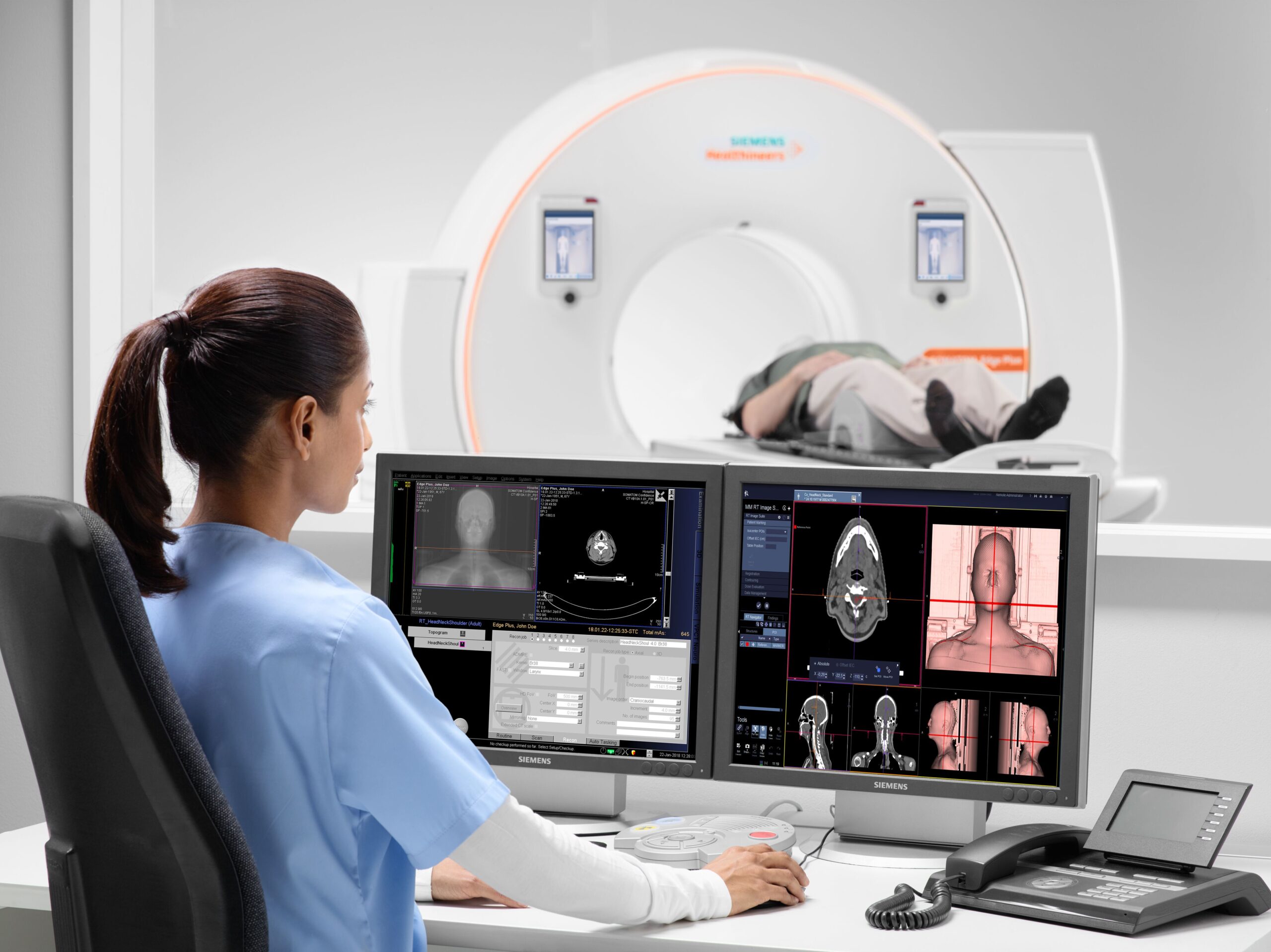
CT devices are movable tables with open ends, around which a tube and detectors rotate. For imaging, you will be positioned on this table. Sometimes pillows, special devices, or securing straps are used to ensure immobility. When positioned correctly, the tube and detectors rotate around you, and the necessary image slices are taken.
Main areas of examination:
- Tumors, fractures, and other diseases of muscles and bones.
- Determining the location and size of tumors, infections, bleeding, and blood clots.
- Guiding for surgical procedures, biopsy, and radiation therapy.
- Diagnosing cancer, heart disease, lung nodules, tumors, and liver masses.
- Evaluating the effectiveness of various treatments, especially cancer treatment.
- Diagnosing internal organ damage and internal bleeding.
Is it risky?
Your scan will be performed by trained and experienced radiology technicians using a computer in the control room. The technician will observe you on a television monitor and communicate with you via intercom during the scan.
Since CT is a more detailed imaging method, you will be exposed to slightly more ionizing radiation than a direct X-ray. While long-term harmful effects of this radiation have not been demonstrated, very high doses have been reported to slightly increase the risk of cancer. However, considering that CT provides much more important information, this risk is considered negligible compared to the potential benefits. New and advanced technology, fast devices are used to perform scans with the lowest possible doses compared to older devices.
There have been no significant adverse effects reported from the low radiation doses used in CT; however, if you are pregnant, your doctor may recommend other diagnostic methods, such as ultrasound (US) or magnetic resonance imaging (MRI), to protect your baby from potential exposure.
In contrast-enhanced (medicated) scans, there may be serious allergic and medical issues, though rare. It is important to inform your doctor if you have a history of allergies.
How to prepare:
- Ask your doctor who requested the CT scan to explain why it is needed, its possible risks, and any questions you may have.
- Depending on the area of the body being examined, you may need to undress and wear a disposable hospital gown provided by the facility.
- You must remove items such as belts, jewelry, dentures, glasses, etc., that could affect the image.
- To obtain clearer images, contrast agents may be used orally, intravenously, or rectally, depending on the area being examined.
- For non-contrast (non-medicated) scans, you may eat, drink, and take your medications. For contrast-enhanced scans, do not eat or drink a few hours before.
- For imaging of infants and children, sedatives (calming medications) may be used to keep the child calm and still. Your doctor will inform you about this.
- If a CT angiogram (CTA) or virtual colonoscopy is requested, additional instructions related to these procedures will be provided when scheduling your appointment.
After the scan:
- Once the scanning procedure is completed, if no special care or attention is needed, you can return to your normal daily life.
- If contrast material was used, you may be observed for a short period until you feel fine.
- The obtained images will be reviewed and interpreted by our specialist doctors and then delivered to you or the requesting doctor.
- Your results and images will be stored digitally in our PACS system for future access.
- Through the PACS system, your images can be shared online with your doctors in other countries for consultation.
- The PACS system minimizes the need for film prints, helping contribute to the sustainability of our world as a film-free center.