Coronary / Cardiac CT Angiography
It is a high-resolution, fast method with a low risk of complications and morbidity. It allows the visualization of some details and the structure of plaques inside vessels, which may not be seen with conventional angiography. For example, it can differentiate between soft plaques, which can lead to sudden deaths due to myocardial infarction, and calcified plaques, as well as determine the degree of stenosis (narrowing). All of these are achieved with very low radiation doses thanks to the latest-generation and most advanced technology devices available in our center.
In CT angiography, it is crucial to capture a moment when the heart rate is at its lowest, which is typically during late diastole. To capture this moment, synchronization with an electrocardiogram (ECG) is required. There are two types of synchronization: prospective and retrospective. The Aquilion One Vision Edition device in our center has the superior feature of prospective ECG synchronization, making this device unique in minimizing radiation dose. Additionally, with the automatic arrhythmia detection software in this device, the use of beta-blockers, which is commonly required in current CT scanners for heart rate control, is no longer mandatory for each patient.
According to published studies in this field, coronary CT angiography performed with such a device has a high sensitivity of about 99%, a positive predictive value of 92%, and a negative predictive value of 95%. With these features, it is a strong alternative to coronary catheter angiography and is progressing towards becoming the new gold standard in diagnosis. Thanks to its ability to exclude coronary artery stenosis (narrowing), it holds an undeniable superiority in patients scheduled for invasive catheter coronary angiography but with a low-to-medium likelihood of coronary artery disease. However, for patients with stenosis that suggests a need for treatment, catheter coronary angiography remains the first choice, as it offers the option of intervention and treatment during the procedure.
While performing coronary CT angiography, the pulmonary arteries, thoracic aorta, and other intra-thoracic structures can also be visualized, allowing for the differential diagnosis of conditions that may mimic acute coronary syndrome. It enables earlier detection of plaques in the vessels and differentiation between life-threatening soft plaques and calcified plaques. It is preferred in patients who have undergone coronary bypass graft surgery or stent placement, as it is non-invasive. It is also a strong option for patients who are unsuitable for cardiac MRI due to reasons like claustrophobia, pacemakers, etc.
For TAVI (transcatheter aortic valve implantation) patients, it is important for determining valve size and identifying the vascular route for catheter advancement. Additionally, 3D (three-dimensional) modeling can be done for this patient group.
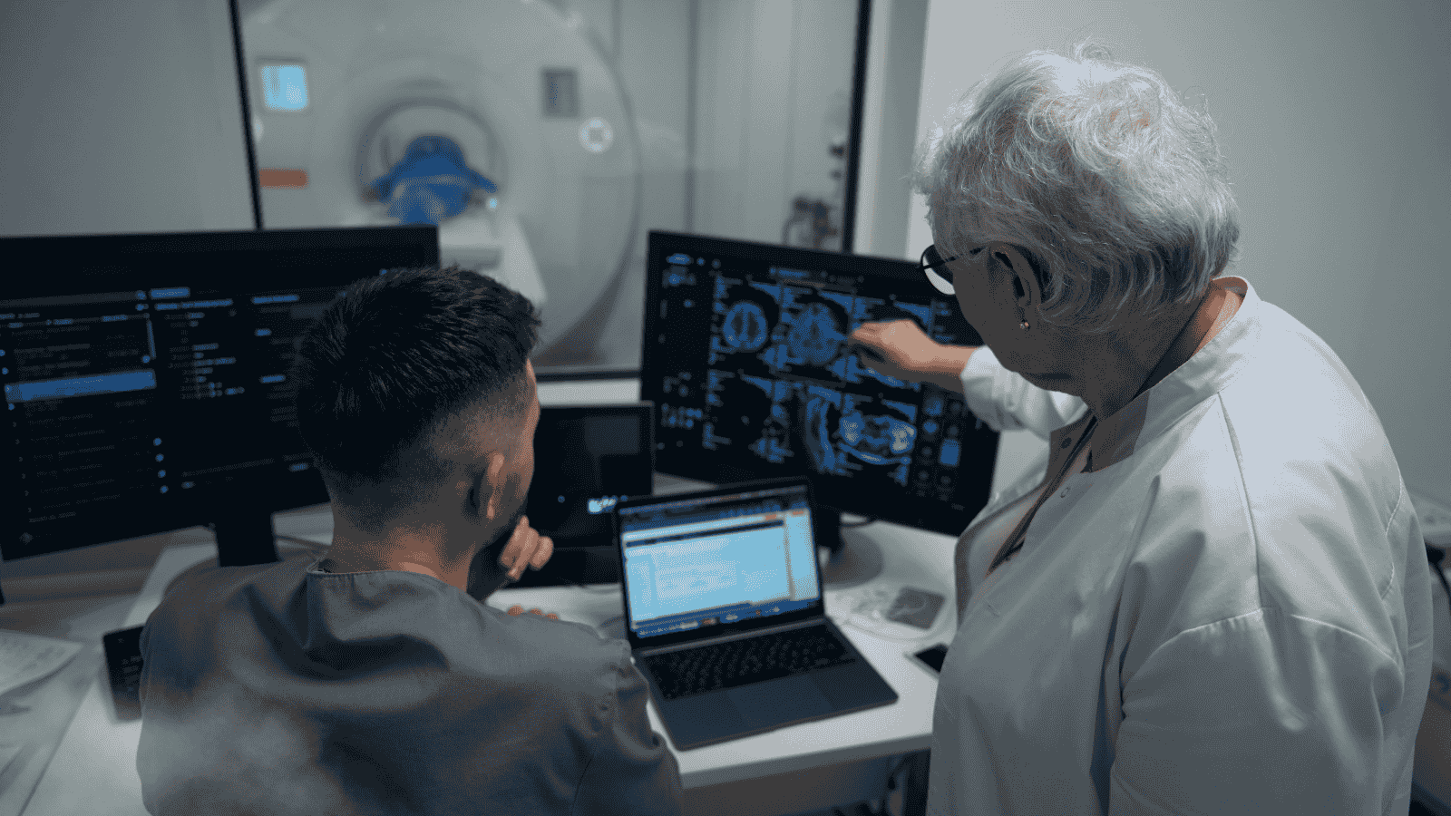
Thanks to specialized and advanced workstation equipment, a large amount of data obtained can be processed, and experienced radiologists can create very detailed reports.
Why is it needed?
- In patients with atypical chest pain
- In diagnosing coronary artery disease
- In patients with ambiguous (unclear) exercise stress test results
- In evaluating coronary bypass grafts
- In detecting stent stenosis (narrowing)
- For imaging the cardiac vein before ablation therapy
- In evaluating abnormal coronary artery anatomy
- In diagnosing congenital heart diseases
- For determining valve size in TAVI patients
- In coronary artery calcium scoring
- In low-dose pediatric exams (chest, coronary angiography, brain, etc.)
Is it risky?
After your preparatory steps, the procedure will be performed by trained and experienced radiology technicians using a computer in the control room. The technician will be able to see you on the monitor and communicate with you via intercom.
Although the low radiation doses used in CT scans have not been shown to have significant adverse effects on humans, if you are pregnant, your doctor may suggest alternative diagnostic methods such as ultrasound (US) or magnetic resonance imaging (MRI) to protect you and your baby from potential exposure.
In contrast (contrast-enhanced) scans, there may be some rare but serious allergic or medical issues. If you have a history of allergies, it is important to inform the requesting or performing physician.
How should we prepare?
First, ask your physician who requested the CT scan to explain why it is being done, the potential risks, and any questions you may have. Depending on the area of your body being examined, you may be asked to undress and wear a disposable gown provided by our facility. You will need to remove any items that may affect the image, such as belts, jewelry, dental prosthetics, glasses, etc.
For clearer images, contrast material may need to be administered orally, through an IV, or rectally (by enema), depending on the area being examined. For non-contrast (non-contrast-enhanced) scans, you may eat, drink, and take your medications as usual. If contrast is required, you will need to refrain from eating and drinking a few hours before the exam.
In pediatric and infant scans, sedatives (calming medications) may be necessary to keep the child calm and still during the procedure. Your physician will inform you about this in advance.
After the scan:
Once the scan is completed, you can return to your normal daily activities unless special care or precautions are required. If contrast material was used, you may receive specific instructions, and you may be monitored briefly until you feel well.
Your images will be reviewed and interpreted by our expert physicians, and a report will be generated. The report will be delivered to you or the requesting physician.
Your results and images will be stored digitally in our PACS system for easy access when needed. Through our PACS system, images can also be shared with your doctors overseas for consultation if requested.