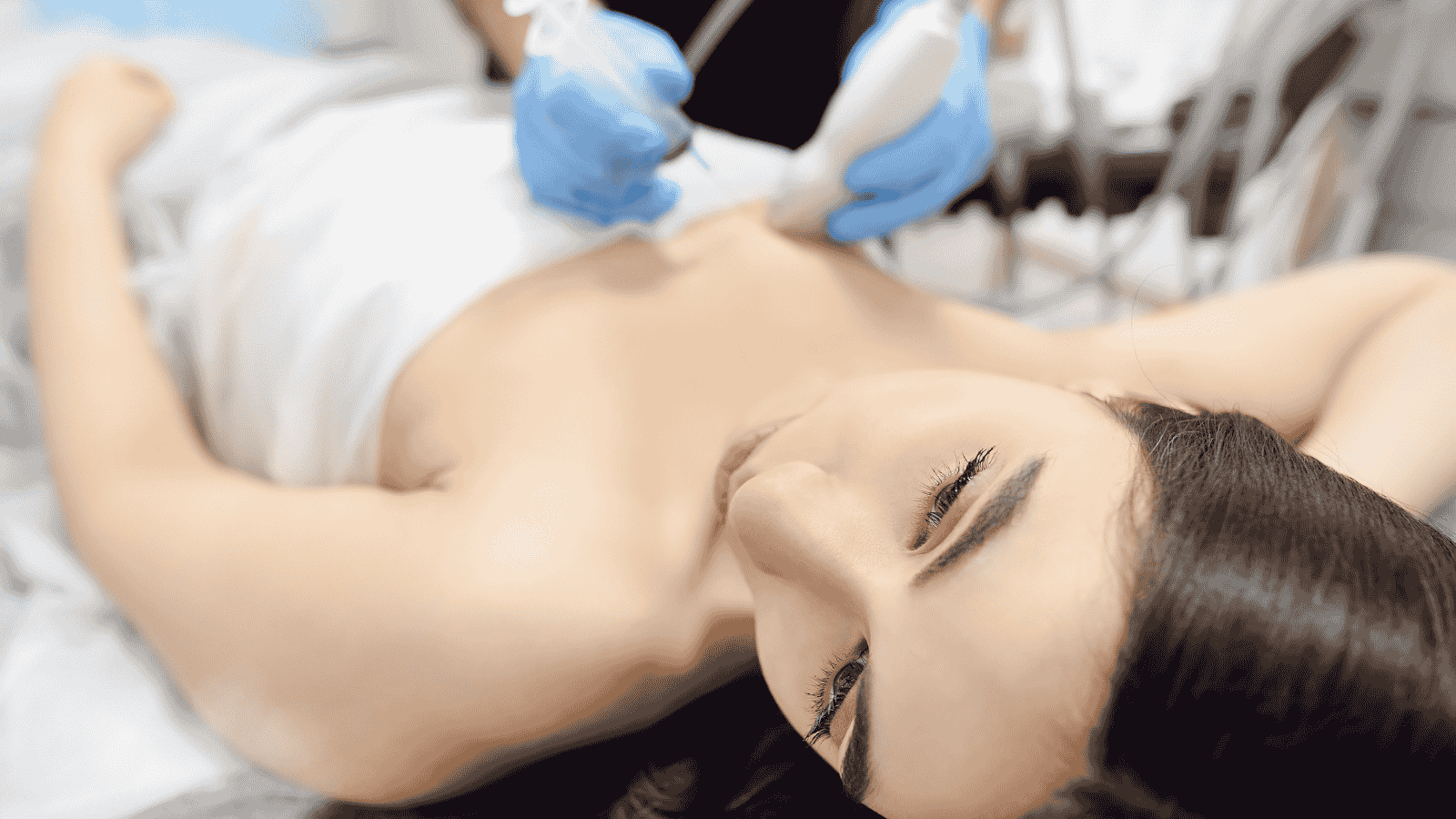
Vacuum-Assisted Breast Biopsy
You will be taken to the procedure room based on the guidance method used. After local anesthesia is applied, a small incision is made at the site where the biopsy needle will be directed to the target tissue. Tissue samples are collected using an automatic mechanism, with approximately 12-24 samples taken, which are then sent to the pathology laboratory for examination. A small metal clip is placed at the lesion site. This procedure, known as marking, serves as a guide for possible future surgical procedures. If surgery is not required, there is no need to remove the clip.
Why is it needed?
This is a highly sensitive method used to determine whether a suspicious mass or lesion in the breast tissue is benign or malignant, and for the differential diagnosis of the cell type required for treatment planning. It is especially prominent in diagnosing cases with calcification or small lesions.
Main areas of examination:
- In the presence of a mass felt during self-examination or by the physician
- In the presence of any suspicious findings such as a mass, lesion, or calcification detected by any imaging method
- In cases where the detected lesions are small, and targeting the exact area is difficult
Is it risky? This interventional biopsy method is a very safe procedure with a low risk of complications. Rare issues such as bleeding, hematoma, or infection may occur. Your physician will take the necessary precautions in this regard.
How should we prepare? This is an outpatient procedure that does not require hospitalization, fasting, or sedation beforehand.
It is important to inform your physician about any existing medical conditions, such as allergies, bleeding disorders, or clotting issues, before the procedure.
Additionally, if you are using anticoagulant medications such as aspirin, clopidogrel, warfarin, or similar drugs, you should pay attention to your physician’s instructions to manage bleeding risks before the appointment.
After the procedure: The incision site will be covered with a bandage. After a short period of observation, you can resume your daily activities. It is not advisable to take a shower until the bandage is removed. Any mild pain or itching at the wound site typically heals within 1-2 weeks.