Ultrasonography (USG)
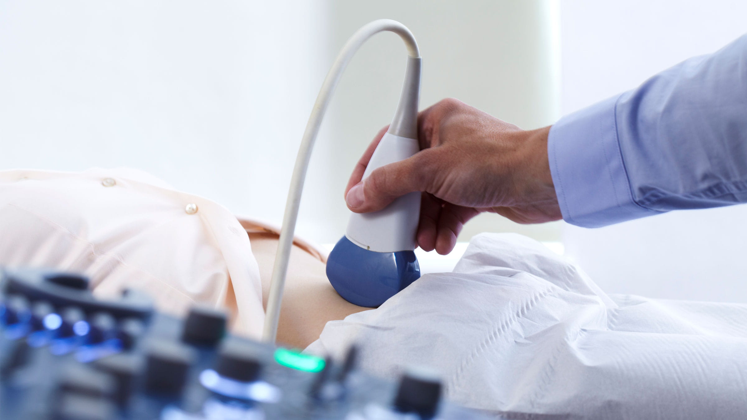
The ultrasound machine consists of a computer console, a monitor, and a transducer, called a probe, that transmits sound waves into the body. During the procedure, you may need to lie on an examination table, sit, or assume different positions as required. The doctor performing the examination will apply a colorless and odorless special gel to the skin of the area being examined to facilitate the transmission of sound. This procedure is painless, tolerable, and may occasionally require you to hold your breath. For certain areas, it may be necessary for your bladder to be full. In this case, the pressure from the probe might cause a feeling of compression. The ultrasound probe is applied to the skin, through the vagina (transvaginal), through the rectum (transrectal), or through the esophagus (transesophageal). The procedure lasts approximately 10-30 minutes. In early pregnancy, an ultrasound examination is performed using a transvaginal probe, and the images can be obtained in 2D, 3D, or 4D (while in motion). As ultrasound is a real-time imaging method, it is also used as a guide for minimally invasive procedures such as needle biopsies or fluid aspirations.
Is it risky?
Prenatal and diagnostic ultrasound is a safe method with no known adverse effects on the baby, mother, or patients.
How to prepare?
This method requires minimal preparation, but for better visualization of certain areas, your bladder may need to be full. If it is not sufficiently full, you will be required to drink water before the procedure. For the examination of some regions and organs, fasting may be expected. This information will be provided in advance when scheduling your appointment.
After the procedure:
After this painless, drug-free, quick, and tolerable procedure, you can immediately receive your report and return to your daily activities.