Cardiac MRI
What is Cardiac MRI?
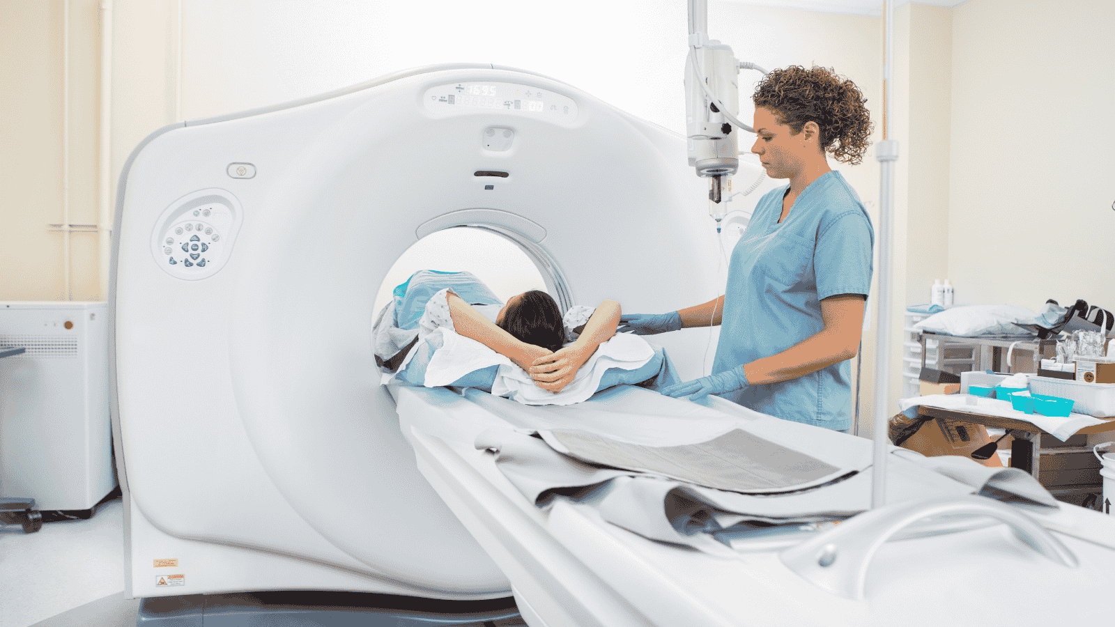
MRI (Magnetic Resonance Imaging) / (3-Tesla MRI) is a non-invasive, painless, and hospitalization-free advanced medical imaging method that provides high-resolution images of the body using a powerful magnetic field, radio waves, and advanced computers with specialized software. Unlike direct X-ray imaging (radiography) and computed tomography (CT), MRI does not use potentially harmful ionizing radiation, making it a patient- and doctor-friendly technique. In the near future, revolutionary advancements are expected with AI-assisted MRI scans, leading to faster imaging, reduced workload, and increased efficiency. Artificial intelligence and machine learning hold promise in areas such as scan planning, multidimensional data acquisition, clinical image interpretation, and personalized imaging.
Cardiac MRI is a safe and non-invasive advanced imaging method used for the diagnosis, treatment, and follow-up of heart diseases. Due to its location in the body and its continuous movement, the heart is one of the most challenging organs to image. However, with the latest high-tech devices (such as 3-Tesla MRI) and specialized software available at our center, these challenges have been overcome, allowing for detailed imaging of heart rhythm, tissue structure, and functions. Since cardiac contraction is assessed in sync with an ECG (electrocardiogram), even a single heartbeat can be captured with clear and high-resolution motion images.
Unlike general MRI scans, cardiac MRI requires the placement of disposable adhesive ECG electrodes on specific areas of the chest. Since heart contractions are evaluated alongside ECG monitoring, high-quality and high-resolution images can be obtained, even within a single heartbeat. For male patients, a small area of the chest may need to be shaved to ensure proper electrode adhesion. These electrodes allow the technician to monitor the heart rate and, when necessary, instruct the patient to hold their breath. This helps prevent motion artifacts that could affect image clarity.
Unlike other imaging methods, MRI is not affected by bones that may obscure or degrade image quality, making it a crucial tool for precise diagnoses.
Additionally, cardiac MRI is a reference method for measuring the volume of heart ventricles and assessing their function.
Why is a Cardiac MRI Needed?
- To evaluate the anatomy and function of the heart chambers, valves, major blood vessels, and pericardium (heart membrane).
- To diagnose and monitor tumors, infections, and inflammatory diseases of the heart and blood vessels.
- To assess the effects of a heart attack and coronary artery disease on heart muscle tissue and to guide treatment planning.
- To determine the causes of heart rhythm disorders (arrhythmias).
- To assist in catheter ablation and other arrhythmia-related procedures.
- To detect pericardial effusion (fluid accumulation in the heart membrane).
- To diagnose and monitor constrictive pericarditis.
- To evaluate heart valve regurgitation (leakage) and stenosis (narrowing).
- To detect heart enlargement (cardiac hypertrophy).
- To diagnose aortic aneurysms and dissections (tears).
- To assess pulmonary hypertension.
- To diagnose, treat, and monitor masses around the heart tissue.
- To track changes in certain heart diseases over time and plan appropriate treatments.
- To diagnose congenital heart and blood vessel abnormalities in both children and adults, assist in surgical planning, and monitor post-surgical outcomes.
For all details regarding MRI (Magnetic Resonance Imaging) / (3-Tesla MRI), preparation, procedures, and advanced MRI applications, click here.